
Clinical Diagnosis and Treatment of Enteropathy-associated T-cell Lymphoma
DOI:
https://doi.org/10.58195/amr.v1i1.13Keywords:
Enteropathy, T-cell lymphoma, clinical diagnosis and treatment analysisAbstract
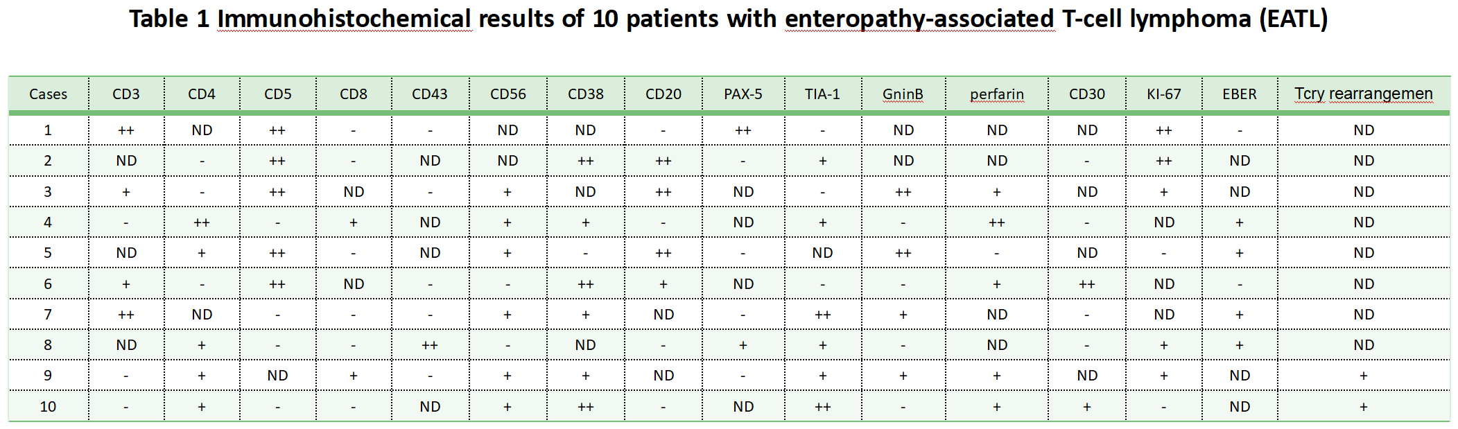
To investigate the clinical characteristics of enteropathy-associated T-cell lymphoma(EATL) and to give treatment strategies. METHODS: From 2019. 02-2022. 02, 10 patients with enteropathy-associated T-cell lymphoma were seen in our hospital, their clinical data were retrospectively analyzed, and practical treatment plans were provided. Results:None of the patients had a history of intestinal disease and all presented to the hospital because of symptoms occurring in the gastrointestinal tract.Five patients had intestinal perforation and underwent dissection, three had small bowel masses and underwent surgery, and two had T-cell lymphoma and underwent related treatment. Six patients had lesions in the small intestine and the others had lesions in the rectum. According to AnnArbor clinical d staging, 9 patients(90%) had stage I+II and 1 patient(10%) had stage III. Gastrointestinal lymphoma AnnArbor clinical staging was the same as lugano clinical staging. 6 patients with B symptoms, 6 with physical status score ≥ 2, 5 with IPI > 2, 6 with PIT > 6; all patients had normal examination regardless of bone marrow examination, β2-microglobulin, or glycoconjugate antigen-125, erythrocyte sedimentation rate. 1 patient with anemia, 6 with decreased albumin, 2 with with elevated lactate dehydrogenase, and 1 case with increased C-reactive protein. According to immunohistochemical hints, 6 cases were CD56 positive and 4 in situ hybridization EBER positive; in the mid-term evaluation of combined patients, the complete remission(CR) rate was 90% in 10 patients; in the DA-EPOCH regimen, the complete remission(CR) rate was 80% in 10 patients. At the completion of 6 chemotherapy courses, all chemotherapy patients were in complete remission(CR) at the end of the period assessment, but 3 of them relapsed within one year, and the disease could not be controlled with the implementation of second-line regimen and chemotherapy again. The follow-up period was 1-25 months, with a high follow-up rate of 100% up to the follow-up time, and 6 patients died, with an overall 2-year survival rate of 40%. Conclusion: Enteropathy-associated T-cell lymphoma is highly aggressive, and after chemotherapy, patients can be treated but are prone to relapse. The use of allogeneic hematopoietic stem cell transplantation(ASCT) becomes the first-line consolidation therapy, which cannot obtain long-term efficacy and needs to be explored.
References
[1] Huang P.Y., Guo X.W., Zuo G.W., et al. A case of monomorphic epitheliophilic intestinal T-cell lymphoma [J]. Journal of clinical gastroenterology. 2018,(2).119-121.
[2] Yu QH, Chen Chaowu. A case of duodenal descending enteropathy-associated T-cell lymphoma causing obstructive jaundice[J]. Chinese Journal of Gastroenterology and Imaging(electronic version). 2018,(4).185-186.
[3] Wang DY, Zhou Q, Wang ZX. The value of gemstone energy spectrum CT small bowel imaging in small bowel lesions[J]. Journal of rare diseases. 2018,(5).56-58.
[4] Wang L, Huang DH, Xiao ChunYan, et al. Report of three cases of monomorphic epitheliophilic intestinal T-cell lymphoma and review of the literature[J]. PLA Medical Journal. 2018,(1).45-50.
[5] Wang BiaoMeng, Zhang Jiucong, Liu DeKe, et al. Report of a case of enteropathic T-cell lymphoma presenting as multiple ulcers in the colon[J]. Journal of gastroenterology and hepatology. 2016,(1).50-52.
[6] Hu YM, Zhang ZY, Liu LY, et al. Clinicopathological analysis of 9 cases of enteropathy-associated T-cell lymphoma[J]. Journal of Clinical and Experimental Pathology. 2015,(6).624-627.
[7] Peng T, Liu JC, Abbas, et al. A case of small intestinal enteropathic T-cell lymphoma[J]. Chinese Journal of Surgical Oncology. 2015, (2).124-125,126.
[8] Xu TJ. One case of type II enteropathy-associated T-cell lymphoma and review of the literature[J]. Journal of rare diseases. 2014,(6).31-33,60.
[9] Zhou J, Shen Q, Ma J, et al. Clinicopathological observation of type II enteropathy-associated T-cell lymphoma[J]. Chinese Journal of Pathology. 2013,(1).26-31.
[10] Ji Liuzhou, Sun Guoyun, Li Hongtao, et al. Value of energy-spectrum CT isotonic mannitol gastrointestinal hypotension angiography for the diagnosis of gastrointestinal diseases[J]. Journal of Medical Imaging. 2013,(12).1948-1952.
[11] Hu YM, Zhang ZY, Liu LY, et al. Clinicopathological analysis of nine cases of enteropathy-associated T-cell lymphoma[J]. Journal of Clinical and Experimental Pathology, 2015,(6):624-627.
[12] Zhou J, Shen Q, Ma J, et al. Clinicopathological observation of type II enteropathy-associated T-cell lymphoma[J]. Chinese Journal of Pathology. 2013,42(1):26-31.
[13] WANG Wenjun, ZHANG Fan(Corresponding author), XU Guoxiang. Clinicopathological observation of type II enteropathy-associated T-cell lymphoma[J]. China Health Care and Nutrition(Midterm Journal). 2013,(6):47-48.
[14] Yang YQ, Wang L, Fan L, et al. Analysis of clinical features of 7 cases of enteropathy-associated T-cell lymphoma[J]. Chinese Journal of Practical Internal Medicine. 2015,35(2):155-158.
[15] Shen Y, Yin Wanyi, Jia Xiaohui, et al. A case of enteropathy-associated T-cell lymphoma and review of the literature[J]. Journal of Doubtful Diseases. 2013,12(6):473-474.
Downloads
Published
How to Cite
Issue
Section
License
Copyright (c) 2022 Yudi Miao, Qiaojiajie Zhao

This work is licensed under a Creative Commons Attribution-NonCommercial-NoDerivatives 4.0 International License.
This is an open-access article distributed under the terms of the Creative Commons Attribution-NonCommerical-NoDerivatives 4.0 International License.


